Dry-Type Macular Degeneration
Drusen
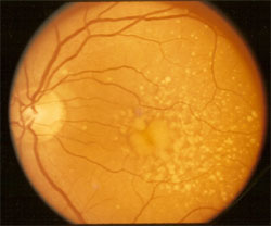
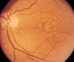
Drusen comprise the classical picture of dry-type macular degeneration. The left eye of the patient CL, shown below, is remarkable for the multiple cream-colored, and slightly elevated soft drusen that have formed deep within the macular and perimacular retina. CL, a Caucasian with blue irides, was 64 years old and her blood pressure was 142/84. The visual acuity was 20/40 for both eyes.
Drusen Regression with Low Salt Diet
Macular degeneration develops
particularly in individuals who use or have used excessive dietary sodium. Several clinical trials have shown, moreover, that macular degeneration develops with greater frequency for people who have a history of mildly elevated blood pressure. The common denominator is that both hypertension and dietary sodium produce an elevated osmotic filtration pressure within the deeper portion of the macular retina.
 |
 |
|
Nervehead is to the left. Numerous cream-colored drusen within the macular and peimacular retina |
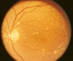
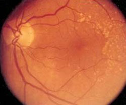
Disappearance of drusen within the central macular following 500 mg sodium diet |
The photo of CL on right (above) shows the improvement that was obtained for the right eye following 25 months adherence to regimented low sodium (500-750 mg daily) and restricted cholesterol diet. The drusen regressed to a remarkable degree and the visual acuity improved to 20/20. The blood pressure improved, too, to the 118/80 level. Blood cholesterol decreased from 238 to 174.
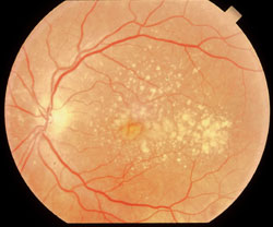
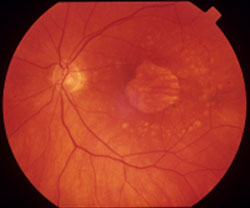
Similar results are shown below for two additional patients who maintained a 500-750 mg sodium restricted diet for 30 months.
 |
 |
|
Small macular drusen. 20/30 acuity |
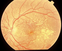
Regression macular
drusen 31 months. 20/20 acuity |
Reversal of soft drusen within the central macular retina can be consistently expected, although sometimes the dietary regiment must be maintained for as long as 42 months before regression occurs. Drusen outside the macular area are less likely to resolve. The latter are not particularly dangerous, however, and their persistence does not threaten retinal integrity.
 |
 |
|
Large, soft drusen 20/30 acuity |
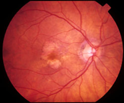
Drusen regression - 22
months. Na restriction 20/20 acuity |
Drusen can be compared with cholesterol plaques that develop within the larger arteries of the body, a process termed atherosclerosis. The coronary, carotid, and renal arteries and the ascending and descending aorta are particularly susceptible to atherosclerosis. For such deposits to develop the blood cholesterol level does not need to be particularly high. What is required is hypertension. The latter, even when the blood pressure elevation is of mild degree only, increases the osmotic pressure across the inner lining of blood vessels with the result that cholesterol, proteins, and serum diffuse into unwanted areas (the wall of an artery).
Atrophy
At the early stage drusen cause little or no visual disability. If regression is not forthcoming by dietary means, the drusen, over a period of many years, cause degeneration (atrophy/death) to the adjacent retinal cells. (see below) At that point flat foci of atrophy become apparent within the macula, and when the degeneration has reached an advanced degree, the foci have a white appearance. These degeneration specks tend to enlarge over time, and when the precise center of the macula becomes involved, visual acuity can decrease to a profound degree.
 |
 |
|
Geographic atrophy
secondary to drusen |
Retinal atrophy, macular
retina |
The process of drusen development, enlargement, regression, and atrophy can be visually disabling even without progressing to the wet stage of the macular degeneration process. For more information about macular degeneration and for particulars regarding the wet-stage complication visit us by clicking here.
|